SWIR Technology
Skin testing is a well-known technique for allergy diagnosis: it is both sensitive and specific. Tests consist of introducing a liquid containing a small amount of potential allergen on the skin and observing the skin’s reaction. If the person is sensitive to the allergen, then a raised inflammatory reaction will occur on the skin called a wheal.
The difficult part is then to measure the size of the wheal reaction on the skin. This lengthy process is usually done manually by a technician and requires advanced training and tedious methods to be accurate enough to provide reliable results.
Traditional visible range color cameras have been used in an attempt to automate and expedite this process but without success. When the skin reacts to an allergen, it typically changes to red, which can sometimes be detected with a standard visible range camera. However, the redness corresponding to an allergic reaction can be diffuse and inconsistent. Moreover, depending on the skin tone, it can be very difficult to detect a change in color caused by this reaction in patients with dark skin color.
Experiments have thus been done with infrared cameras to try to better detect this reaction regardless of skin tone or skin color pigmentation. However, the specific type of infrared wavelength has an impact on the utility of this approach. Typically, thermal infrared cameras (LWIR) do not provide adequate detection of an allergic reaction on the skin as they do not detect the wheal very well.
Flare Diagnostics has tested and incorporated the use of Short-Wave Infrared (SWIR) cameras sensitive between 0.9 and 1.7 µm for this detection. The picture below illustrates SWIR’s ability to get a clearer and well contrasted signature of an allergen skin test compared to other technologies. Flare’s patent pending approach outperforms visible range, near infrared (NIR) and thermal range (LWIR) for detecting the wheal.


Experiments have also been performed with SWIR technology on various skin tones to determine the utility of this approach when skin pigmentation may make allergen induced wheal detection more difficult. Figure 2 demonstrates SWIR imaging of several allergy test on 8 subjects, 2 of which have darker skin tone. This helps illustrate that SWIR is agnostic to skin color, providing clear detection of an allergic reaction on the skin regardless of skin pigmentation. These images would not be possible using visible range or other technologies evaluated.
When a proper filter is utilized with the SWIR camera, strong contrast of the wheal versus surrounding skin is apparent regardless of skin tone. Moreover, SWIR imaged skin test reactions are very clear and sharp, providing the possibility of a more automated approach to detection. Combined with artificial intelligence (AI), this automated approach has the potential to increase throughput and safety compared to traditional methods.
SWIR cameras with InGaAs detectors are sensitive between 0.9 and 1.7 µm. In this spectral band, water absorbs lights making droplets easily detectable.
SWIR cameras are thus powerful tools to detect moisture and waterdrops.
This can be seen on the following picture.

Figure 3: Picture of water dropping on a surface, obtained with a Bobcat 320 camera from Xenics.
Flare Diagnostics proprietary method is to use SWIR camera ability to detect water as a means to resolve allergic reactions on the skin. Indeed, allergen induced wheal reactions are due in part to a small amount of liquid, mainly water, located under the skin.
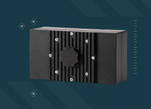
In the Flare Diagnostics skin test detection approach, the SWIR camera currently utilized in practice is a Bobcat 320 from Xenics. This camera is based on a 2D focal plane array of 320 x 256 pixels with a 20 µm pitch. It is a small and light camera, 55 x 55 x 72 mm3 and only 285 g, thus very easy to implement on a global system. Moreover, thanks to its intuitive Xeneth graphic user interface (GUI) it is easy to use or integrate into a larger system.

Figure 4: SWIR camera currently utilized in practice is a Bobcat 320.
Flare Diagnostics’ proprietary AllergyScope system consists of the SWIR camera and speciality designed neural net software that can be used to capture skin test images and automatically perform a detailed measure of the size of a wheal reaction. Traditional measurement techniques require manual and individual measurement of the spots using a ruler. This is both time consuming, error prone and difficult to integrate into electronic medical records. The AllergyScope camera system from Flare Diagnostics on the other hand, acquires a global picture of the skin and thanks to the strong contrast in the SWIR range, it can instantaneously, accurately and automatically document the wheal pattern with high precision. Allergy skin testing is thus made faster, easier, and potentially more accurate with the AllergyScope. Moreover, these results are consistent regardless or skin color or tone.
The AllergyScope system from Flare Diagnostics is currently based on a 320 x 256 resolution camera, however Xenics also offers SWIR cameras in advanced 640 x 512 resolution as well. For future systems it is thus possible to get even more precise and sensitive skin test measurements. Xenics’ new generation Wildcat camera offers a real improvement in terms of resolution (four times the number of pixels compared to a 320 x 256 camera) and sensitivity (30% better). Wildcat is very compact (55 x 55 x 72 mm3 in CameraLink version) and is available in a plug-and-play configuration with a USB3 interface.

Figure 5: Xenics’ new generation Wildcat camera.
Thanks to Flare Diagnostics’ AllergyScope system and highly sensitive SWIR cameras from Xenics, it is now possible to have fast and accurate allergic diagnostic imaging, giving more flexibility to medical teams and simplifying the process for the patients. Moreover, the latest state of the art SWIR technologies currently available help pave the way for the next generation of systems with better sensitivity and thus potentially more accurate diagnostics.
AUTHORS:
Jody R. Tversky
Assistant Professor of Medicine
Johns Hopkins University School of Medicine
Marc Larive
Strategic Marketing Manager
Xenics nv
Documentation
146370


Related content
Apr 09th 2024
Exosens Adds Innovative Photon Counting System, LINCam

Feb 06th 2024
Occar renew their trust in Photonis' expertise
Photonis shall deliver 40,000 image intensifier tubes to boost military night vision in Germany and Belgium

KINTEX Exhibition Center Ⅱ.
FROM Sep 25th 2024 TO Sep 28th 2024
DX KOREA 2024
Visit Exosens at DX KOREA 2024 from September 25 to 28 in KINTEX Exhibition Center Ⅱ, Korea

Shanghai New International Expo Centre.
FROM Jul 08th 2024 TO Jul 10th 2024
Vision China 2024
Visit Exosens at Vision China 2024 from July 8 to 10 in Shanghai New International Expo Centre, China

Paris Nord Villepinte exhibition centre.
FROM Jun 17th 2024 TO Jun 21st 2024
Eurosatory 2024
Visit Exosens at Eurosatory 2024 from June 17 to 21 in Paris Nord Villepinte exhibition centre, France

Berlin.
FROM Jun 05th 2024 TO Jun 09th 2024
ILA Berlin 2024
Visit Exosens at ILA Berlin 2024 from June 5 to 9 in Berlin, Germany.

Anaheim Convention Center, Anaheim, California.
FROM Jun 02nd 2024 TO Jun 06th 2024
ASMS 2024
Visit Exosens at ASMS 2024 from 2 to 6 June 2024 at the Anaheim Convention Center, Anaheim, California, USA.
National Harbor, Maryland.
FROM Apr 21st 2024 TO Apr 25th 2024
SPIE Defense & Commercial Sensing 2024
Visit Exosens at SPIE Defense & Commercial Sensing 2024 from 9 to 12 April 2024 in National Harbor, Maryland, USA.

Munich.
FROM Mar 09th 2024 TO Apr 12th 2024
Analytica 2024
Visit Exosens at Analytica 2024 from 9 to 12 April 2024 in Munich, Germany